Myocardial segmentation using contour-constrained optical flow tracking
Despite the important role of object tracking using the Optical Flow (OF) in computer graphics applications, it has a limited role in segmenting speckle-free medical images such as magnetic resonance images of the heart. In this work, we propose a novel solution of the OF equation that allows incorporating additional constraints of the shape of the segmented object. We formulate a cost function that include the OF constraint in addition to myocardial contour properties such as smoothness and elasticity. The method is totally different from the common naïve combination of OF estimation within
Myocardium segmentation in strain-encoded (SENC) magnetic resonance images using graph-cuts
Evaluation of cardiac functions using Strain Encoded (SENC) magnetic resonance (MR) imaging is a powerful tool for imaging the deformation of left and right ventricles. However, automated analysis of SENC images is hindered due to the low signal-to-noise ratio SENC images. In this work, the authors propose a method to segment the left and right ventricles myocardium simultaneously in SENC-MR short-axis images. In addition, myocardium seed points are automatically selected using skeletonisation algorithm and used as hard constraints for the graph-cut optimization algorithm. The method is based
In silico design and experimental validation of sirnas targeting conserved regions of multiple hepatitis c virus genotypes
RNA interference (RNAi) is a post-transcriptional gene silencing mechanism that mediates the sequence-specific degradation of targeted RNA and thus provides a tremendous opportunity for development of oligonucleotide-based drugs. Here, we report on the design and validation of small interfering RNAs (siRNAs) targeting highly conserved regions of the hepatitis C virus (HCV) genome. To aim for therapeutic applications by optimizing the RNAi efficacy and reducing potential side effects, we considered different factors such as target RNA variations, thermodynamics and accessibility of the siRNA
Improved estimation of the cardiac global function using combined long and short axis MRI images of the heart
Background: Estimating the left ventricular (LV) volumes at the different cardiac phases is necessary for evaluating the cardiac global function. In cardiac magnetic resonance imaging, accurate estimation of the LV volumes requires the processing a relatively large number of parallel short-axis cross-sectional images of the LV (typically from 9 to 12). Nevertheless, it is inevitable sometimes to estimate the volume from a small number of cross-sectional images, which can lead to a significant reduction of the volume estimation accuracy. This usually encountered when a number of cross-sectional
Inherent fat cancellation in complementary spatial modulation of magnetization
An efficient fat suppression method is presented for MR tagging with complementary spatial modulation of magnetization (CSPAMM). In this method, the complementary modulation is applied to the water content of the tissues, while in-phase modulation is applied to the fat content. Therefore, during image reconstruction, the subtraction of the acquired images increases the tagging contrast of the water while cancels the tagging lines of the fat. Compared with the existing fat suppression techniques, the proposed method allows imaging with higher temporal resolution and shorter echo-time without
Improved technique to detect the infarction in delayed enhancement image using k-mean method
Cardiac magnetic resonance (CMR) imaging is an important technique for cardiac diagnosis. Measuring the scar in myocardium is important to cardiologists to assess the viability of the heart. Delayed enhancement (DE) images are acquired after about 10 minutes following injecting the patient with contrast agent so the infracted region appears brighter than its surroundings. A common method to segment the infarction from DE images is based on intensity Thresholding. This technique performed poorly for detecting small infarcts in noisy images. In this work we aim to identify the best threshold
Improved strain measuring using fast strain-encoded cardiac MR
The strain encoding (SENC) technique encodes regional strain of the heart into the acquired MR images and produces two images with two different tunings so that longitudinal strain, on the short-axis view, or circumferential strain on the long-axis view, are measured. Interleaving acquisition is used to shorten the acquisition time of the two tuned images by 50%, but it suffers from errors in the strain calculations due to inter-tunings motion of the heart, which is the motion between two successive acquisitions. In this work, a method is proposed to correct for the inter-tunings motion by
In-silico development and assessment of a Kalman filter motor decoder for prosthetic hand control
Up to 50% of amputees abandon their prostheses, partly due to rapid degradation of the control systems, which require frequent recalibration. The goal of this study was to develop a Kalman filter-based approach to decoding motoneuron activity to identify movement kinematics and thereby provide stable, long-term, accurate, real-time decoding. The Kalman filter-based decoder was examined via biologically varied datasets generated from a high-fidelity computational model of the spinal motoneuron pool. The estimated movement kinematics controlled a simulated MuJoCo prosthetic hand. This clear-box

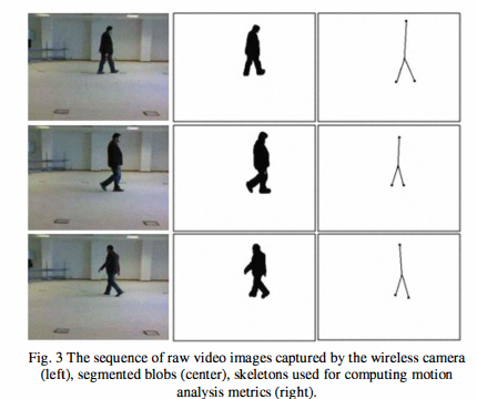
Ambient and wearable sensing for gait classification in pervasive healthcare environments
Pervasive healthcare environments provide an effective solution for monitoring the wellbeing of the elderly where the general trend of an increasingly ageing population has placed significant burdens on current healthcare systems. An important pervasive healthcare system functionality is patient motion analysis where gait information can be used to detect walking behavior abnormalities that may indicate the onset of adverse health problems, for quantifying post-operative recovery, and to observe the progression of neurodegenerative diseases. The development of accurate motion analysis models

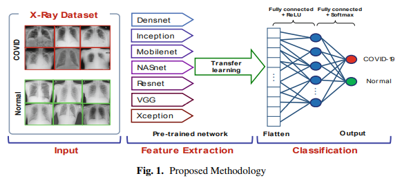
Detection of COVID-19 from Chest X-Ray Images Using Deep Neural Network with Fine-Tuning Approach
The coronavirus (COVID-2019) quickly spread throughout the world and came to be a pandemic. To avoid further spreading this epidemic and treat affected patients rapidly, it is important to recognize the positive cases as early as possible. In this paper, deep learning techniques are employed to detect COVID-19 from chest X-ray images quickly. The images of the two classes, COVID and No-findings are collected from three public datasets. The proposed approach consists of two phases; transfer learning and fine-tuning. Transfer learning is carried out by seven deep learning models: DenseNet
Pagination
- Previous page ‹‹
- Page 10
- Next page ››
