Breadcrumb

Segmentation of the right ventricle in MRI images using a dual active shape model
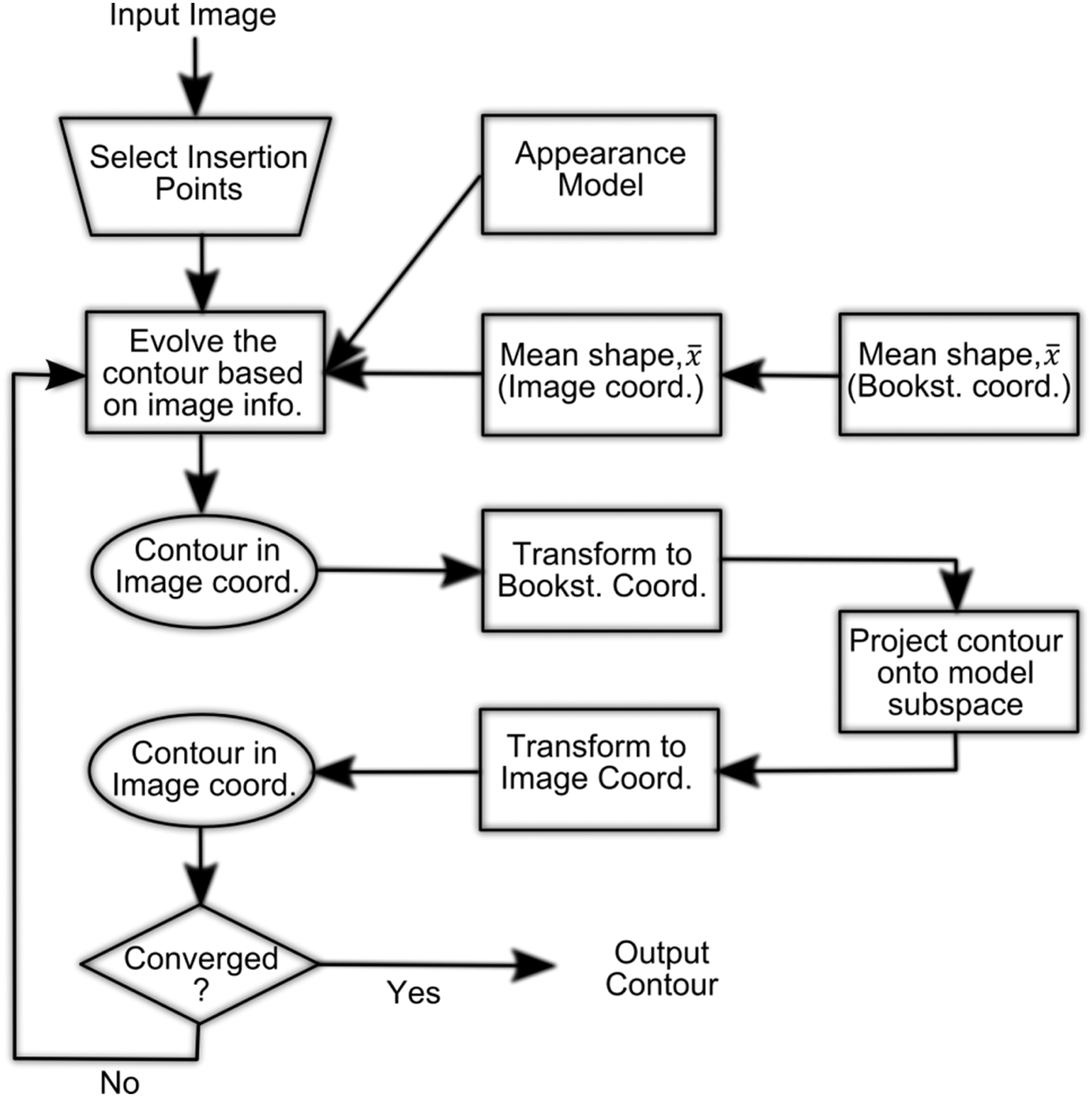
Active shape models (ASM) showed to have potential for segmenting the right ventricle (RV) in cardiac magnetic resonance images (MRIs). Nevertheless, the large variability and complexity of the RV shape do not allow for concisely capturing all possible shape variations among patients and anatomical cross-sections. Noticeably, the latter increases the number of iterations required to converge to a proper solution and reduces the segmentation accuracy. In this study, the authors propose a new ASM framework that can model the RV shape in short-axis cardiac MRI images. In this framework, the RV contour is split into two simpler segments, septal (SP) and free wall, whose shape variations are independently modelled using two separate (dual) ASM models. The contour splitting is done at the location of the RV insertion points into the SP wall. Further, instead of using the conventional Procrustes method, the RV contours are aligned using the Bookstein coordinate transformation, which uses the RV insertion points as landmarks to linearly align the RV contours. The results from a dataset of 14 patients show that the proposed framework outperforms the conventional ASM framework and can model complex RV shape variation with more accuracy and in less iteration steps. © The Institution of Engineering and Technology 2016.


